The medical sector, especially the pathology, is one of our fields of interest. Hyperspectral imaging allows the development of new techniques, such as new methods for the diagnosis of cancer to give an example. This enables faster and cheaper diagnoses, speeding up surgery and cutting down the time the patient needs to be in anesthesia while waiting for the result of the pathologist.
Since our cameras cannot be calibrated before the optics is mounted, the adaption to a microscope was possible but rather complicated. The camera needed to be mounted to the microscope and the final calibration had to be conducted on-site, which means that the conditions for the calibration usually were not perfect.
Development of a relay optics
By developing a relay lens for the hyperspectral FireflEYE, we are able to overcome that disadvantage. The camera and the relay lens are assembled in our premises, where it directly gets calibrated according to our high quality standards.
With the relay lens extension, we provide full flexibility to our customers as they are able to directly work with the camera (plug-and-play) – whether it shall be mounted on a microscope, be used with one of our common lenses with different focal lengths, or even be attached to any special optics with C-mount.
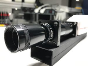
First prototype of the relay lens attached to the FireflEYE
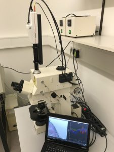
FireflEYE 185 attached to a Zeiss Axiophot
In order to demonstrate how easy it is to work with the camera we attached the relay lens to the C-mount adapter of a Zeiss Axiophot, which was provided by the ILM (Institute of Laser Technologies in Medicine and Measurement Technique), a research institute in Ulm. From the beginning, i.e. mounting the camera to the microscope, until the first measurement it only took a few minutes and we received a sharp image with a richness of detail.
Getting reflectance values is even easier than in any other measurement environment. By simply using the microscope in light transmission mode instead of having the light source illuminating the sample from above, the camera can be white-calibrated by taking an image without a sample placed on it. Since the light shines through the sample, the transmitted signal carries the relevant information because of particular absorption features within the sample. By this, we achieve great results with a necessary integration time of not more than 0.1ms.
Histological analysis
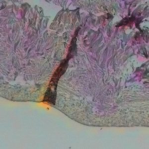
We analyzed a histological slice of pig skin that was cultured on a chicken egg. Under the microscope the sample shows a complex structure, which occurs in different colors. The figure below shows a colored infrared (CIR) image of the pigskin histology on the left-hand side, while on the right-hand side the spectral reflectance of all pixels inside the corresponding rectangles with the same color in the image can be seen. Since the Axiophot has a NIR filter for wavelengths larger than 750nm the spectral range is limited to this value.
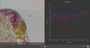
Colored Infrared Image (CIR), histological slice of pigskin. Purple: Collagen and reticular network, Yellow: Erythrocytes, Grey/pink: Cytoplasma and muscle tissue
Visualizing hidden details
The different colors occur because of the dyeing methods that were applied to help visualizing different features of the histological sample: coagulated areas because of the laser cut, healthy skin, hair follicles, blood vessels, erythrocytes.
Analyzing the hyperspectral image of the samples allows the user to extract hidden features, visualize those by different band settings in a RGB output and even quantifying the results automatically, e.g. with the help of machine learning. See below some more examples of the histological measurement taken with the hyperspectral FireflEYE 185 .
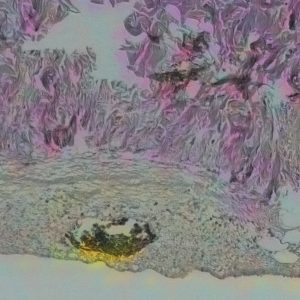
Hair follicle (CIR)
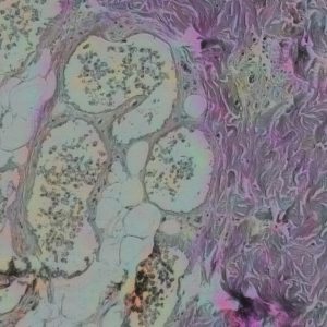
Healthy skin cells with supply vessels and cell nuclei (CIR)
Collagen and reticular network in purple, cytoplasma and muscle tissue in grey/pink, blood vessel with erythrocytes in yellow (CIR)
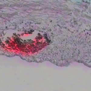
The same blood vessel and erythrocytes, displayed in another CIR band combination and magnified
